Image Credits
The following banner and highlight images are used on the IMC21 website. They have been submitted to the RMS either directly or through our biennial scientific imaging competition.
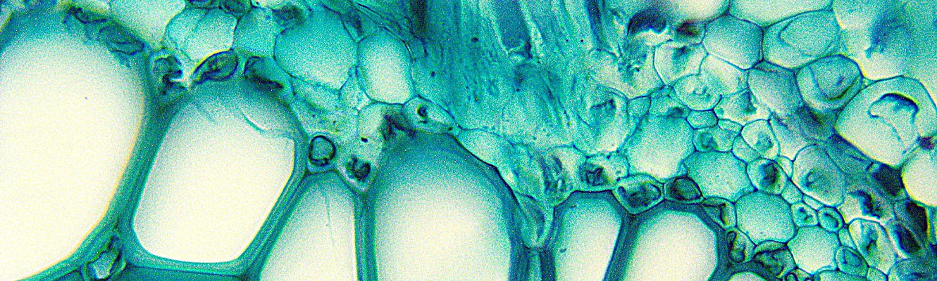
Fern Stem - Fern stem cross-section under the microscope, Magnification 400x .Science botany micrograph plant root tissue cells. Microscope used: AmScope T340 Series Trinocular Compound Microscope.
Viorica Calota
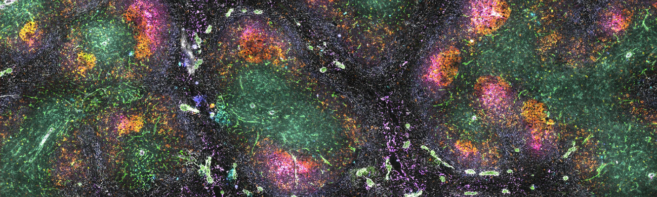
Spleen galaxy - The spleen is an organ of the lymphatic system that plays an important role in the immune system. In this image, multiple markers were used to characterize the structure of the germinal centres in the spleen. 25 different cellular markers were targeted including B cells, T cells, plasma cells, fibroblasts, macrophages, stromal cells, granulocytes, red blood cells and dendritic cells.
Isabel San Martin Molina
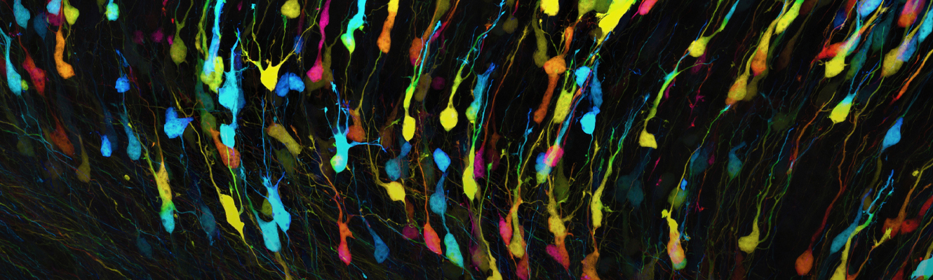
Neurons 1: Depth-coded projection of neurons within the cerebral cortex of the developing brain - This composite image of neurons within the cerebral cortex of the brain was generated by Dr Anthony J. Hayes on a Zeiss LSM880 confocal microscope using a 63x/1.4 plan-apochromat oil immersion objective . It was assembled from 120 separate image stacks, each stack comprising 46 optical sections. The tissue volume sampled was approximately 1mm x 1.8mm x 20µm which took two and a half hours in total to capture.
The image reveals the complex neuronal circuitry of the brain. Tissue depth is represented by colour (deep structures shown in cold colours e.g. shades of blue; superficial structures in warmer colours, e.g. yellows, oranges and reds).
Tissue sample was kindly provided by Dr Isabel Martinez Garay.
Anthony J. Hayes
A Colorized Nanoscale Portrait with Graphene Oxide - False-colored image of graphene oxide flake which was taken by a FEI Titan TEM. The structure resembles a woman’s profile.
Xuyun Guo
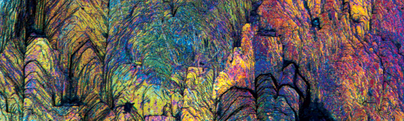
Oxidized Town - The notion of microcosm in Greek philosophy compares our world with the universe (the macrocosm). The artistic/scientific image capture the microcosm observed on the surface of a pyrite mineral specimen, to reveal a world that exists around us but which the human eye cannot see. The idea is to question the notion of the universe, scales of size and microcosm. On the scale of the universe, we ourselves are microscopic, insignificant and fragile.
This approach articulates practice and theory through the relationship between science, art and society, in order to develop a mechanism and a junction between these themes that can be anchored in the issues of contemporary visual arts.
The photomicrographed subject was the oxidized surface of pyrite mineral, using a Zeiss Axioskop microscope with 5x magnification and DIC technique.
Jonathan Muyal
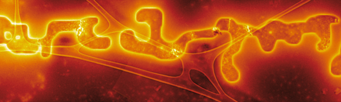
Mysterious Message - Colourised bright-field transmission electron microscopy image montage of metal-chloride negative stain on apoferritin. The brightly outlined regions are a crystalline sodium chloride drying artefact which strikingly resembles illegible writing, and reminds us we don't always see what we expect to under the microscope.
Benjamin L Weare