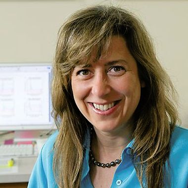
Congress Symposia
IMC21 will consist of three congress symposia comprising 32 individual sessions. Please click the down arrows for more information of each session. Please note, this information is subject to change.
Abstract submission is now open for talk and poster presentations. The deadline for submissions is Friday 6 March 2026.
Instrumentation
Innovating Microscopy End-to-End: Hardware, Automation, and AI in Honour of Albert Crewe
Session Chairs: Ondrej Krivanek (USA), Paul Midgley (UK)
This symposium showcases innovations across the electron microscopy pipeline - from advanced electron optics and novel components (e.g., aberration correctors, phase plates) to automated acquisition and intelligent data analysis. It covers both real-time (on-the-fly) and conventional offline processing, highlighting advances in machine learning and AI for image enhancement, reconstruction, and quantitative interpretation. By integrating cutting-edge hardware with smart software, the symposium promotes interdisciplinary collaboration and presents tools that enhance accessibility, reproducibility, and new imaging modalities in materials science, life sciences, and beyond. Contributions from diverse fields that bridge instrumentation, automation, and data science are warmly encouraged.
Invited Speakers
Diffraction and 4D-STEM
Session Chairs: Jian-Min Zuo (Australia), Colin Ophus (USA)
This symposium highlights advances in electron diffraction-based techniques, spanning methodology and applications in electron crystallography. It covers converged beam (CBED), precession, and electron backscattered diffraction (EBSD), along with scanning-based diffraction imaging via 4D-STEM dataset. Emphasis is on novel instrumentation, data acquisition, and computational approaches enabling nanoscale phase identification, strain and symmetry analysis, structure solution, electromagnetic field mapping, and more. The session invites contributions that advance quantitative diffraction techniques and demonstrate impactful applications across materials science, chemistry, and structural biology.
Invited Speakers
Dynamical, In-situ & Environmental Microscopy
Session Chairs: Eva Olsson (Sweden), Utkur Mirsaidov (Singapore)
This symposium explores development in dynamic, in‑situ, and environmental microscopy, especially those that capture materials and processes under real‑world conditions. Topics include ultrafast microscopy, with and without ultrafast lasers, as well as in‑situ studies of samples in diverse physicochemical environments: ultra‑high vacuum, controlled gas atmospheres (from low to ambient pressure), liquid cells, and under dynamic stimuli such as electrical biasing, light illumination, temperature extremes (cryogenic to high‑T), and mechanical stress. Emphasis is placed on operando techniques that emulate functional conditions in energy, catalysis, electronics, and biological systems. Contributions showcasing innovative experimental designs, instrumentation, and insights into time‑resolved structural or chemical evolution are especially encouraged.
Invited Speakers
Advances in Phase Imaging Techniques
Session Chairs: David Muller (USA) and Vincenzo Grillo (Italy)
This symposium explores recent advances in phase imaging across electron and photon microscopy, with a focus on quantitative methods that recover phase information for nanoscale structure and field mapping. Topics include ptychography (with electrons and X-rays), off-axis and in-line holography, Lorentz microscopy for magnetic and electric field visualization, and emerging techniques employing structured electron or photon probes (such as vortex beams, tailored wavefronts, and coherence-engineered illumination) to enhance sensitivity and resolution. The session welcomes contributions on algorithmic innovations, instrumentation, and applications in materials science, quantum systems, and life sciences that leverage phase contrast for unprecedented insight into electric, magnetic, and strain fields at the nanoscale.
Invited Speakers
Quantitative and multimodal 3D imaging in the Physical Sciences
Session Chairs: John Miao (USA)
This symposium will highlight advances in quantitative and multimodal 3D imaging that reveal the structure, composition, and properties of materials across length scales, from individual atoms to functional devices. It will feature cutting-edge tomography and phase-retrieval methods, including atomic electron tomography (AET/pAET), multislice electron ptychography, ptychographic X-ray nano/micro-tomography, atom probe tomography (APT), and volume electron microscopy (e.g., FIB-SEM, serial block-face SEM). Emphasis will be placed on quantitative reconstruction, multimodal data fusion, and AI-driven interpretation, integrating imaging, spectroscopy, and theory to yield physically validated 3D and 4D datasets. The session will also focus on correlative workflows, cross-modality integration, and standardized data protocols that promote reproducibility and interoperability. Applications from materials science, nanotechnology, energy, catalysis, and geoscience are encouraged, particularly those advancing predictive modelling and design through FAIR, AI-ready 3D data.
Indicative topics (not limited to):
• Atomic electron tomography (AET) and ptychographic AET (pAET)
• Multislice electron ptychography and 4D-STEM phase retrieval
• Ptychographic X-ray nano/micro-tomography
• Correlative multimodal tomography (tomography + EDX/EELS, holography, CDI/ptychography)
• Volume EM, FIB-SEM, and X-ray tomography across length scales
• Advances in atom probe tomography (APT) and TEM–APT correlative workflows
• AI for 3D/4D imaging: reconstruction, segmentation, registration, and uncertainty quantification
• Quantitative metrology, calibration standards, data reproducibility, and FAIR data practices
Invited Speakers
Surface sensitive microscopy, analysis and sample manipulation (SEM, FIB, Helium microscopy and LEEMS and PEEMS etc).
Session Chairs: Wenxin Tang (China)
This symposium highlights advances in surface-sensitive imaging and analysis techniques, as well as methods that engineer near-surface regions through etching, ablation, or additive approaches. It welcomes contributions on scanning electron microscopy (SEM), focused ion beam (FIB) (used for both nanoscale modification and imaging), helium ion microscopy (HIM), secondary ion mass spectrometry (SIMS), and low-energy electron microscopy (LEEM/PEEM). Emphasis is on new instrumentation, in situ capabilities, correlative workflows, and enhanced surface sensitivity. Applications in catalysis, 2D materials, thin films, semiconductors, and energy materials are encouraged, especially those combining characterization with precise surface engineering.
Invited Speakers
Scanning probe microscopy and advanced light microscopy
Session Chairs: Sorin Stanescu (UK) and Ioanna Mela (UK)
This symposium brings together innovations in scanning probe microscopy (SPM) and advanced light microscopy, highlighting synergies and complementary advances in nanoscale imaging, spectroscopy, and manipulation. Topics include high-resolution atomic force microscopy (AFM), scanning tunnelling microscopy (STM), near-field optical techniques (e.g., SNOM, nano-FTIR), super-resolution fluorescence microscopy, light-sheet imaging, and correlative multimodal approaches. Emphasis is placed on novel probe designs, functional imaging under realistic conditions (e.g., in liquid, at cryogenic or elevated temperatures), quantitative nanomechanical/electrical/optical mapping, and integration with other analytical platforms. The symposium welcomes contributions that push the limits of spatial, temporal, and spectral resolution, and that demonstrate impactful applications across physics, materials science, biology, and nanotechnology.
Invited Speakers
Advanced in Spectroscopy and Spectral Imaging Techniques
Session Chairs: Demie Kepaptsoglou (UK) and Donald MacLaren (UK)
This symposium highlights recent advances in electron-based spectroscopy and spectral imaging, including innovations in electron energy-loss spectroscopy (EELS), energy-dispersive X-ray spectroscopy (EDX), cathodoluminescence (CL), and electron-beam-induced current (EBIC). It also welcomes contributions on the emerging techniques that exploit coherent coupling between electron beams and laser fields, enabling new forms of photon-induced near-field electron microscopy (PINEM), ultrafast spectroscopies, and quantum-coherent probes. The focus is on instrumental developments, data quantification methods, and multimodal correlative approaches that enhance spatial, spectral, and temporal resolution. Contributions demonstrating novel applications in materials characterization, nano photonics, quantum materials, and life sciences are warmly welcomed.
Invited Speakers
Coming soon...
Life Sciences
Light Microscopy - Live Imaging Cells, Tissues, Organs and Organisms
Session Chairs: Kurt Anderson (UK) and Helen Ishikawa-Ankerhold (Germany)
This symposium will focus on recent developments in live imaging that enable the observation of biological processes within their native environments. Topics will include the use of fundamental methods through to advances in light-sheet and lattice microscopy, adaptive optics, and high-speed volumetric techniques that preserve physiological conditions while achieving enhanced spatial and temporal resolution. Presentations will also address innovations in labelling, environmental control, and image analysis that support quantitative interpretation of dynamic biological systems. The session will provide an overview of how contemporary live imaging approaches are enabling detailed study of cellular and tissue function across multiple scales.
Invited Speakers
Light Microscopy - Resolution Revolution
Session Chairs: Ralf Jungmann (Germany), Ilaria Testa (Sweden)
This symposium will highlight the continuing evolution of light microscopy techniques that overcome the diffraction limit to achieve nanometre-scale resolution. Presentations will describe theoretical and technological advances in methods such as STED, SIM, single-molecule localisation, MINFLUX, and expansion microscopy. The session will examine improvements in optical design, probe chemistry, and computational reconstruction that enhance precision and reliability, and will include the integration of these techniques into quantitative workflows for structural and molecular analysis. Together, these developments illustrate the ongoing refinement of light microscopy as a tool for high-resolution visualisation of complex biological systems.
Invited Speakers
Coming soon...
Light Microscopy - Volumetric Imaging
Session Chairs: Joerg Bewersdorf (USA), Amanda Foust (UK)
This symposium will examine methods that extend light microscopy into three or more dimensions for the detailed reconstruction of complex biological specimens. Topics will include light-sheet and multi-view imaging, adaptive optics, remote focusing, and advanced computational approaches for image registration and deconvolution. Presentations will consider the technical challenges associated with imaging thick and scattering samples, including strategies for optical clearing, refractive index matching, and labelling uniformity. The session will present the latest capabilities in volumetric data acquisition and analysis, highlighting progress towards comprehensive three-dimensional characterisation of biological structure and dynamics.
Invited Speakers
Light Microscopy - Democratisation and Widening Access to Microscopy
Session Chairs: Richard Bowman (UK) and Jan Huisken (Germany)
This symposium will address initiatives that broaden access to microscopy technologies, expertise, and data. Presentations will describe efforts in open-source instrument design, low-cost optical systems, community-based facilities, and digital training resources. The session will also consider approaches to reproducibility, standardisation, and sustainable operation in diverse research environments. Emphasis will be placed on frameworks that promote equitable access, capacity building, and long-term technical support. Collectively, these contributions reflect ongoing work to ensure that advances in microscopy are accessible to a wide and inclusive global research community.
Invited Speakers
Multimodal Imaging - Plant Sciences
Session Chairs: George Littlejohn (UK), Mark Fricker (UK)
This symposium will explore developments in multimodal imaging applied to plant biology. Presentations will focus on the integration of optical, spectroscopic, and electron-based techniques to examine plant structure and function across scales. Topics will include imaging approaches for cellular and subcellular organisation, tissue architecture, and whole-organ analysis. The session will also consider methodological advances that address the unique optical and structural challenges posed by plant specimens. Through the combination of complementary modalities, multimodal imaging continues to enhance understanding of plant development, physiology, and environmental interaction.
Invited Speakers
Coming soon...
Multimodal Imaging - Neurosciences
Session Chairs: Michael Grange (UK)
This symposium will focus on the application of multimodal imaging to the study of neural structure and function. Presentations will describe recent progress in combining light, electron, and spectroscopic techniques to investigate the organisation of neural circuits and the molecular composition of brain tissue. Topics will include correlative workflows, registration methods, and computational integration across spatial scales. The session will highlight methodological developments that facilitate the linkage of structural and functional information, providing a comprehensive view of the nervous system across organisational levels.
Invited Speakers
Multimodal Imaging - Imaging Pathogen Biology from Structure to Mechanism
Session Chairs: Dan Hurdiss (Netherlands) and Shee-Mei Lok (Singapore)
This symposium will present recent advances in multimodal imaging of pathogens and host–pathogen interactions. Topics will include the combination of cryo-electron microscopy, tomography, and advanced light microscopy with analytical and structural methods to elucidate infection processes at molecular and cellular resolution. Presentations will address imaging across temperature, spatial, and temporal regimes, together with approaches for sample preservation, correlation, and quantitative interpretation. The session will provide an overview of how integrated imaging techniques are enabling detailed examination of pathogen structure and mechanism.
Invited Speakers
Coming soon...
Multimodal Imaging - Correlated Imaging Across Length Scales
Session Chairs: Anna Kotowska (UK)
This symposium will address methodologies that link imaging data across spatial and temporal scales to create coherent, multiscale representations of complex systems. Presentations will describe advances in correlative light, X-ray, and electron microscopy, as well as integration with analytical and spectroscopic techniques. Topics will include registration strategies, fiducial systems, and data alignment frameworks that maintain spatial accuracy between modalities. The session will outline emerging solutions for managing and analysing large, multimodal datasets and will cover the role of correlated imaging in bridging molecular, cellular, and macroscopic perspectives.
Invited Speakers
Enabling Technologies - Machine Learning in Biological Imaging
Session Chairs: Juliette Griffié (Sweden)
This symposium will focus on the growing role of machine learning in the acquisition, reconstruction, and interpretation of biological image data. Presentations will address algorithmic developments in segmentation, denoising, and object recognition, as well as machine learning–driven approaches to adaptive imaging and automated analysis. Topics will include network architectures, training strategies, validation, and reproducibility within quantitative imaging workflows. The session will also consider the implementation of artificial intelligence within microscopy facilities and its impact on data quality, throughput, and discovery across the life sciences.
Invited Speakers
Coming soon...
Electron Microscopy - Cryo-ET - Macromolecular imaging in cells, organoids and tissues
Session Chairs: Elizabeth Wright (USA) and Jurgen Plitzko (Germany)
This symposium will highlight the latest developments and applications of cryogenic electron tomography (cryo-ET) for three-dimensional imaging and in situ structural analysis of biological processes in cells, organoids, and tissues. Presentations will cover recent advances in sample preparation, instrumentation, and data acquisition strategies that improve both resolution and throughput. Speakers will also discuss cutting-edge computational approaches for segmentation, identification, and 3D reconstruction of macromolecular complexes within the crowded cellular environment. The symposium will showcase how these innovations are driving new biological discoveries, providing molecular insights into cellular organisation and host–pathogen interactions. Finally, the session will consider future perspectives for macromolecular imaging at the cell and tissue scale, highlighting both the challenges and opportunities ahead. It will explore how advances in structural systems biology can bridge the gap between atomic-level understanding of individual macromolecules and the emergent functions they collectively drive within cells, tissues, and whole organisms.
Invited Speakers
Electron Microscopy - Cryo-EM - Innovations in macromolecular structure determination
Session Chairs: Arjen Jakobi (Netherlands) and Rouslan Efremov (Belgium)
This symposium will explore recent breakthroughs in cryogenic electron microscopy (Cryo-EM) that are transforming our understanding of macromolecular architecture and dynamics. The session will highlight advances in microscope and detector technology, image processing algorithms, and sample preparation methods. Talks will showcase how these innovations enable structure determination of complex biomolecular assemblies, transient conformations, and membrane-bound proteins. Looking ahead, the symposium will also examine how Cryo-EM interfaces with emerging computational approaches, particularly protein structure prediction and machine learning. The integration of Cryo-EM with AI-driven modelling tools such as AlphaFold and advanced image analysis pipelines is reshaping structural biology (accelerating model building), improving map interpretation, and guiding experimental design. Together, these innovations promise to expand the reach of Cryo-EM toward dynamic, data-driven understanding of macromolecular function in health and disease.
Invited Speakers
Coming soon...
Electron Microscopy - Volume imaging
Session Chairs: Lucy Collinson (UK) and Leandro Lemgruber (UK)
This symposium highlights advances in volume electron microscopy (vEM) that are transforming how we study the three-dimensional organization of biological systems. Presentations will cover cutting-edge methods such as serial block-face and focused ion beam scanning electron microscopy (SBF-SEM and FIB-SEM), array tomography, and correlative light and electron microscopy (CLEM). These approaches enable high-resolution mapping of cells, tissues, and organ systems in unprecedented detail, revealing structural relationships that underpin biological function. Speakers will discuss innovations in sample preparation, imaging automation, and computational reconstruction that make large-scale, quantitative 3D imaging increasingly accessible. By showcasing applications ranging from neuroscience and cell biology to developmental and host–pathogen interactions, this symposium will demonstrate how vEM is providing a new window into the complex architecture of life.
Invited Speakers
Multimodal imaging - Biomineralisation
Session Chairs: Michael Elbaum (Israel) and Fabio Nudelman (UK)
Session information coming soon...
Invited Speakers
Physical Sciences
Soft materials, polymers, pharmaceuticals, and beam sensitive materials
Session Chairs: Xiaodong Zou (Sweeden) and Sonia Antoranz Contera (UK)
This symposium focuses on the unique challenges and innovative microscopy approaches used to study soft materials, polymers, pharmaceuticals, and other beam-sensitive systems. These materials often exhibit complex, amorphous, semi-crystalline and/or dynamic structures that are critical to their function but are highly susceptible to damage under conventional imaging conditions.
We invite contributions that showcase advanced imaging techniques such as cryo-electron microscopy (cryo-EM), X-ray methods, low-dose TEM/STEM, electron diffraction and 4D-STEM methods, environmental SEM, atomic force microscopy (AFM), and correlative microscopy methods (designed to preserve structural integrity while providing high-resolution insights). Studies on the nanoscale morphology or chemistry, phase behaviour, crystallinity, molecular organization, and interfaces in soft matter systems, polymer blends, drug formulations, and biologically relevant materials are especially encouraged.
Emphasis will be placed on sample preparation strategies, dose minimization, in situ and operando microscopy, and emerging tools for quantitative analysis. Contributions that integrate microscopy with complementary techniques or modeling to unravel structure (property relationships in these delicate materials are welcome).
Invited Speakers
Two-Dimensional Materials
Session Chairs: Juan Carlos Idrobo (USA) and Ute Kaiser (Germany)
This symposium will explore cutting-edge developments in the characterization and analysis of two-dimensional (2D) materials using advanced microscopy techniques. As 2D materials such as graphene, transition metal dichalcogenides (TMDs), and hexagonal boron nitride continue to drive innovations in electronics, photonics, and energy storage, high-resolution imaging and analytical tools are essential for understanding their structural, chemical, and electronic properties at the atomic scale.
Presentations will highlight recent breakthroughs in transmission electron microscopy, scanning electron microscopy, and scanning probe techniques for investigation of topics such as layer stacking, point and extended defects, strain, and interfacial phenomena in 2D materials and 2D heterostructures. Novel 2D materials such as magnetic and ferroelectric structures, twisted 2D heterostructures and encapsulated 2D structures will be discussed. Novel techniques applied to 2D materials will be presented including low dose methods, variable voltage and cryo-TEM to investigate air-sensitive samples. Correlative methods combining microscopies and spectroscopy as well as advanced sample preparation strategies, and data analysis techniques tailored to the unique challenges of 2D systems. The session will cover disciplines across materials science, chemistry, nanotechnology, and microscopy, seeking insight into both fundamental properties and applied aspects of 2D materials.
Invited Speakers
Functional ceramics and oxides
Session Chairs: Maria Varela (USA) and Miaofang Chi (USA)
This symposium will present work demonstrating the power of microscopy to unravel the structure (property relationships in functional ceramics), and complex oxides (materials central to applications in electronics, energy, sensing, and more). Across length-scales from atomic to microscale, the session seeks contributions that illuminate how microstructure, defects, interfaces, and composition govern performance.
A particular topic of interest is functional ceramics engineered for active roles such as ferroelectrics, piezo-electrics, dielectric, electro-optic, magnetic, and multiferroic ceramics. The session will encompass a wide range of advanced microscopy techniques like TEM, STEM, electron diffraction, SEM (including environmental and cryogenic modes), electron tomography, and scanning probe microscopy, applied to reveal domain structure, grain boundary behaviour, defect distributions, phase transitions, and strain fields. Submissions involving thin films, bulk ceramics, composites, coatings, and ceramics under stimulus (electric, thermal, mechanical) are encouraged.
Complex oxide systems of interest include perovskites and spinel's to layered oxides and heterostructures exhibiting functional behaviour (e.g. conductivity, magnetism, catalysis, ion transport). Key microscopy-driven investigations are particularly welcome including use of in situ and operando imaging, high resolution mapping of chemical states (via EELS, STEM-EDS), tracking dynamic phenomena like oxidation/reduction, oxygen vacancy migration, phase evolution, and interface effects. Integration of microscopy with complementary characterization (diffraction, spectroscopy), advanced data analysis and modelling to link nanoscale observations with macroscopic functionality is highly encouraged.
Invited Speakers
Structural materials, metals, and alloys
Session Chairs: Laure Bourgeois (Australia) and Randi Holmestad (Norway)
As demand grows for materials that combine strength, durability, lightweight character, and high-performance under extreme conditions, microscopy is essential for understanding the relationships between structure, defects, and properties. The session will explore the application of advanced microscopy to reveal the microstructural origins of mechanical, thermal, and corrosion-related behaviour in structural metals and alloys. It will present microscopy and spectroscopy tools and insights, that are being applied for advancing the next generation of structural materials, metals and alloys.
Topics include, but are not limited to: grain- and phase-boundary characterization; dislocation networks and stacking faults; precipitates and secondary phase evolution; nano-scale segregation and solute clustering; deformation mechanisms under load; and damage accumulation—cracking, fatigue, creep, oxidation. Presentations using all microscopy methods are most welcome including SEM, EBSD, TEM, STEM, scanning diffraction methods, X-ray computational tomography, optical and acoustic microscopy and spectroscopy. In situ microscopy (mechanical, thermal, environmental), high-resolution imaging and tomography for 3D reconstructions, and correlative microscopy-spectroscopy methods are especially welcome. Contributions that link microstructural observations with alloy design, processing histories, and performance in service will be of particular interest.
Invited Speaker
Nanomaterials and Catalysts
Session Chairs: Qian Chen (USA) and Marc Willenger (Germany)
This symposium will focus on the application of microscopy techniques to understand and engineer nanomaterials and catalytic systems. Nanostructured materials play a central role in a broad range of fields ranging from electronics to energy conversion. Microscopy is an essential tool to uncover structural, chemical, and functional properties across multiple length scales.
Microscopy enables the study of the intricate features of nanomaterials, including morphology, crystallinity, defects, and interfaces. This session will highlight imaging and analytical methods such as scanning electron microscopy (SEM), high-resolution transmission electron microscopy (TEM), scanning TEM (STEM), electron tomography, atomic force microscopy (AFM) and other scanning probe methods applied to investigate nanomaterials including nanoparticles, nanowires, and quantum dots. Emphasis will be placed on advances that uncover structure-function relationships, correlative methods, three-dimensional characterisation, in situ imaging and spectroscopy at high resolution, reducing imaging and characterisation artefacts, as well as data interpretation that enable improved visualization of electronic behaviour and atomic-scale features. Contributions are encouraged that demonstrate how microscopy enables the design and optimization of nanomaterials for electronic, photonic, or mechanical applications.
Catalysts often rely on complex nanoscale architectures where structure and chemistry evolve under operating conditions. Part of this session will therefore focus on catalytic materials, including how in situ and operando microscopy techniques (such as environmental TEM and environmental-cells) are used to study catalytic behavior in real time. Key topics include the identification of active sites, structure-performance relationships, monitoring of dynamic structural changes, and understanding of support of metal interactions. Presentations are encouraged that link atomic-scale observations to catalytic performance, degradation mechanisms, or reaction pathways.
Invited Speakers
Earth and planetary materials
Session Chairs: Christian Vollmer (Germany) and John Wheeler (UK)
This symposium will focus on the characterization of Earth and planetary materials using advanced microscopy techniques to uncover their structural, chemical, and isotopic signatures. From the deep Earth, to cultural heritage, to distant planetary bodies, microscopy plays a critical role in deciphering the formation, transformation, and history of materials, minerals, rocks, meteorites, and extraterrestrial samples at the nanoscale.
We invite contributions that utilize electron microscopy (TEM, SEM, STEM), focused ion beam (FIB) techniques, atom probe tomography, X-ray computed tomography, X-ray spectroscopy and correlative methods to investigate mineral structures, inclusions, zonation, amorphization, shock effects, and fluid/mineral interactions. Special emphasis will be placed on studies of high-pressure phases, presolar grains, returned mission samples (e.g., from the Moon, Mars, asteroids), materials of historical cultural significance, and materials relevant to geochemical cycles or planetary differentiation. In situ and cryo-microscopy approaches that preserve delicate or volatile components are also encouraged.
By bringing together microscopists, geoscientists, and planetary scientists, this session aims to highlight how state-of-the-art microscopy is reshaping our understanding of cultural heritage and planetary processes from atomic-scale mechanisms to our significant historical records or planetary evolution.
Invited Speakers
Magnetic behaviour and Ferroelectrics
Session Chairs: Naoya Shibata (Japan) and Xuiliang Ma (China)
This symposium will focus on the application of advanced microscopy techniques to investigate magnetic and ferroelectric materials, with an emphasis on understanding domain structures, switching mechanisms, and nanoscale interactions that underpin functional behavior. These materials are critical for emerging technologies in memory, sensing, spintronics, and energy conversion.
Contributions are invited that utilize high-resolution imaging methods such as Lorentz TEM, electron holography, canning transmission electron microscopy (STEM) combined with EELS or differential phase contrast imaging, magnetic force microscopy (MFM) and piezoresponse force microscopy (PFM). Particular interest lies in the visualization and manipulation of magnetic and ferroelectric domains, vortex structures, skyrmions, and domain wall dynamics under applied fields, elevated or cryogenic temperature, or as a result of applied strain. Operando and in situ microscopy approaches that provide real-time insight into switching behavior, fatigue, and aging mechanisms are encouraged. Studies addressing thin films, multilayers, and heterostructures (including coupling effects between electric, magnetic, and structural order parameters) are especially welcome.
Invited Speakers
Semiconductors, Quantum and ICT
Session Chairs: Rebecca Nicholls (UK) and David Smith (USA)
This symposium highlights the critical role of advanced microscopy in the development and optimization of materials for semiconductors, quantum technologies, and information and communication technology (ICT) devices. As device architectures become increasingly complex and materials are pushed to atomic-scale limits, microscopy provides essential insights into structure, property relationships that govern performance and reliability.
We welcome contributions that utilize electron microscopy (TEM, STEM, EELS, tomography), scanning probe techniques (STM, AFM), advanced optical and X-ray spectroscopy and correlative or in situ approaches to characterize defects, interfaces, strain, composition, and electronic structure in semiconductors and low-dimensional systems. Topics include, but are not limited to, advanced CMOS materials, wide bandgap semiconductors, semiconductor materials for logic and memory, and quantum materials such as superconductors, topological insulators, and single-photon emitters.
Special emphasis will be placed on imaging challenges in ultra-scaled devices, beam-sensitive materials, and cryo- or operando techniques for observing devices in functional states. Studies linking microscopy with device performance, fabrication processes, or quantum behavior are particularly encouraged.
The session aims to showcase researchers working at the intersection of microscopy, materials science, and device engineering to advance the frontiers of modern electronics and quantum technologies.
Invited Speakers
Energy Storage and Conversion
Session Chairs: Chongmin Wang (USA) and Jordi Arbiol (Spain)
This symposium will focus on the application of advanced microscopy techniques to investigate materials and interfaces critical to energy storage and conversion technologies. As global demand increases for efficient, durable, and sustainable energy systems, including batteries, fuel cells, supercapacitors, and solar cells, microscopy plays a vital role in revealing the structural, chemical, and electrochemical processes that govern performance and degradation.
We invite contributions that employ electron and scanning probe microscopies (TEM, STEM, SEM, AFM, STM), X-ray spectroscopy and microscopy methods, spectroscopic imaging (EELS,
EDS, SIMS), and in situ/operando methods to study materials across multiple length scales. Key topics include phase transformations, solid-electrolyte interphases (SEI), ion transport mechanisms, advanced membrane structures, nanostructured electrodes, electrode evolution, and interface stability during cycling or under operating conditions.
Contributions are welcome that present time-resolved and environmental microscopy, cryo-techniques for sensitive materials, and multimodal or correlative approaches that link microstructural dynamics to electrochemical performance. Submissions bridging experimental observations with modeling or device-level behavior are also encouraged.
The session aims to demonstrate how microscopy is being applied to accelerate the development of high-performance materials for sustainable energy technologies.
Invited Speakers
Radiation Damage and Nuclear Energy: Microscopy at Extreme Conditions
Session Chairs: Jonathan Hinks (UK) and Chris Russo (UK)
This symposium will highlight the use of advanced microscopy to investigate radiation effects in materials including those relevant to nuclear energy systems. Understanding how materials respond to high-energy particle irradiation, through defect formation, deformation, phase transformations, and microstructural evolution, is essential for reliable interpretation of advanced microscopy data as well as for the design of safe, durable nuclear fuels, structural components, and waste forms.
We invite contributions that quantitatively consider structural and chemical evolution during electron, ion or X-ray irradiation. Submissions that integrate microscopy with modelling or correlative characterisation methods are encouraged. We also wish to showcase defect investigations that utilize techniques such as electron microscopy (TEM, STEM, SEM), atom probe tomography, and X-ray methods to characterize irradiation-induced defects such as dislocation loops, voids, bubbles, and segregation phenomena. Studies of materials exposed to ion, neutron, or electron irradiation (both ex situ and using in situ ion irradiation capabilities) are of particular interest. Emphasis will also be placed on imaging and quantifying radiation effects in advanced alloys, ceramics, composites, and coating systems for fission and fusion environments.
Submissions that combine microscopy with atomistic modeling, mechanical testing, or real-time performance evaluation are encouraged, as are studies of radiation tolerance mechanisms and the development of radiation-resistant materials.
The session aims to showcase collaboration between microscopists, nuclear materials scientists, and engineers working to understand and mitigate radiation damage in extreme environments.
Invited Speakers
Phase Transformations and High-Speed Dynamics: Real-Time Microscopy of Evolving Materials
Session Chairs: Renske van der Veen (Netherlands) and William Bryan (UK)
This symposium will explore the use of advanced microscopy techniques to investigate phase transformations and rapid dynamic processes in materials across a range of environments and timescales. Understanding how materials evolve under stimuli such as temperature, pressure, stress, or irradiation is key to tailoring properties for high-performance applications in aerospace, energy, electronics, and more.
We welcome contributions that employ in situ and operando microscopy, particularly transmission electron microscopy (TEM), scanning electron microscopy (SEM), and scanning probe methods, to capture real-time structural, chemical, or morphological changes during transformations such as crystallization, melting, amorphization, grain growth, martensitic transitions, or diffusion-driven reactions. Studies utilizing ultrafast or high-frame-rate imaging, pump-probe techniques, or dynamic TEM (DTEM) are especially encouraged.
Topics may include solid–solid phase transitions, interface migration, defect dynamics, shock responses, and nanoscale mechanisms driving rapid transformation pathways. Submissions that combine experimental imaging with simulations or data-driven analysis to interpret high-speed processes are also welcome.
This session aims to bring together researchers pushing the limits of temporal and spatial resolution in microscopy to better understand the fast, complex phenomena that govern material behavior under extreme or transient conditions.
Invited Speakers
Coming soon...
Last updated: 22/11/2025


